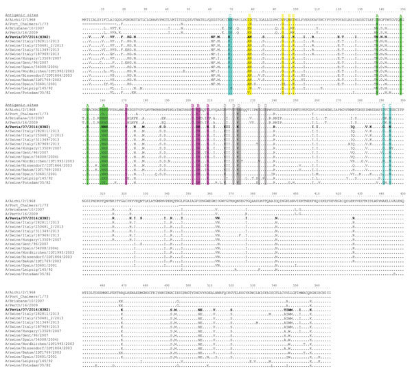
Figure 2.

Amino acid sequence alignment of the hemagglutinin protein of swine influenza virus (SIV) (H3N2) strain A/Pavia/07/2014 (bold) and other SIVs. Antigenic sites A, B, C, D, and E of H3 HA are highlighted in green, magenta, blue, gray, and yellow, respectively, as proposed by others (11). Amino acid changes with respect to the A/Aichi/2/1968 strain are indicated for each strain.
