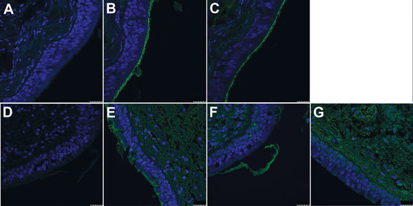
Figure 2.

Interaction of hemagglutinin (HA) of H3-P99 (panels B and E) and H10-JD346 (panels C, F, and G) isolates of influenza A(H10N8) viruses with human trachea. Sections from 2 persons are shown (A–C and D–G). A and D, negative control staining (secondary antibody without HA). Blue indicates nuclei stained with 4',6-diamidino-2-phenylindole; green indicates HA binding. Scale bars indicate 25 μm.
