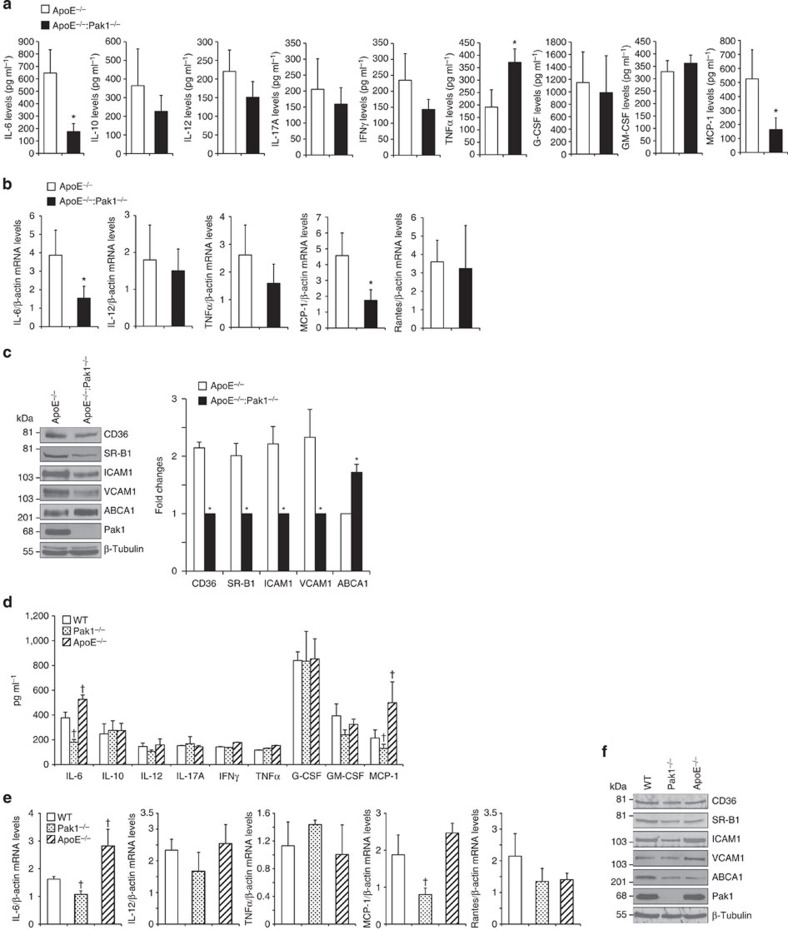
Figure 3. Lack of Pak1 downregulates IL-6 and MCP-1 levels in ApoE−/− mice.
(a) The plasma from ApoE−/− and ApoE−/−:Pak1−/− mice fed with WD for 16 weeks were analysed for the indicated inflammatory cytokines and MCP-1 levels. (b) RNA was isolated from aortic arch region of ApoE−/− and ApoE−/−:Pak1−/− mice fed with WD for 16 weeks and qRT–PCR analysis for the indicated cytokines and chemokines was performed. (c) An equal amount of protein from the aortic extracts of the mice described in a was analysed by western blotting for the indicated proteins using their specific antibodies. Bar graph in c represents the quantification of three western blottings each from a group of two pooled arteries. (d) The plasma from WT, Pak1−/− and ApoE−/− mice fed with CD were analysed for the indicated inflammatory cytokines and MCP-1 levels. (e) RNA was isolated from aortic arch region of the mice described in d and qRT–PCR analysis was performed for the indicated cytokines and chemokines. (f) An equal amount of protein from the aortic extracts of the mice described in d was analysed by western blotting for the indicated proteins using their specific antibodies. Data were presented as mean±s.d. and assessed by Student's t-test. *P<0.01 versus ApoE−/− mice (n=6 mice); †P<0.05 versus WT mice (n=6).

