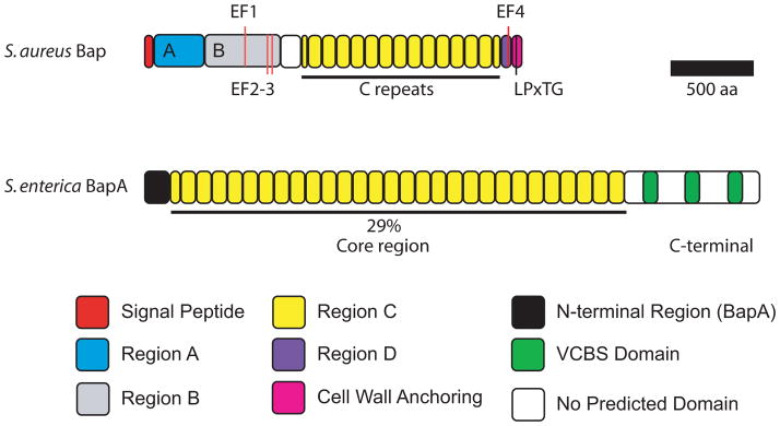
Figure 3.

Domain organization of S. aureus Bap and S. enterica BapA. EF-hand calcium-binding motifs EF1 to 4 in Bap are indicated. LPxTG is the cell-wall anchoring motif. The repeats in the core regions of S. enterica BapA shares 29% identity with the C repeats of S. aureus Bap.
