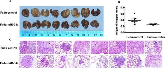
Figure 3. MiR-34a reduced metastatic potential of HNSCC cells in vivo.

(A) The macroscopy of metastasis nodes induced by Fadu-miR-34a and Fadu-control in the lung of nude mice. (B) The weights of mice lungs with metastasis nodes induced by Fadu-miR-34a and control cells (p < 0.01). (C) The histopathology of metastases induced by Fadu-miR-34a and Fadu-control in lung tissues with HE staining (Original magnifications × 100). *necrotic area.
