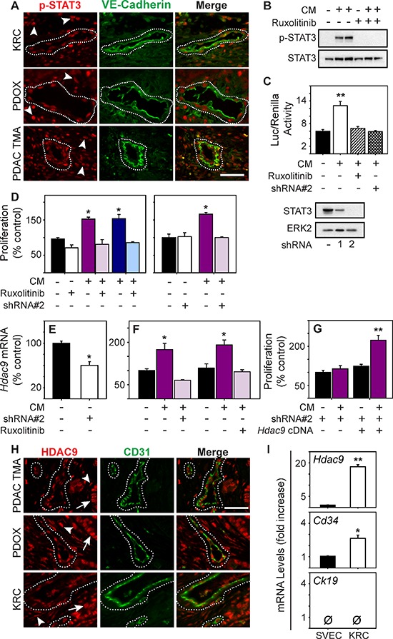
Figure 4. STAT3 is active in PDAC tumor endothelia and enhances HDAC9 expression to promote endothelial proliferation.

(A) p-STAT3 (red) is abundant in the nuclei of VE-Cadherin-positive vessels (green, outlined) and surrounding stromal cells (arrowheads) in KRC (top), EUS-PDOX tumors (middle), and human PDACs (bottom). (B) KRC CM markedly increases p-STAT3 levels in ECs, which is blocked by ruxolitinib [100 nM]. (C) KRC CM significantly enhances STAT3 luciferase reporter activity in ECs (top), which is blocked by ruxolitinib [100 nM] or a STAT3-targeting shRNA (shRNA#2). Immunoblotting (lower panel) shows the knockdown efficiency of STAT3-targeting shRNAs. ERK2 confirms equivalent lane loading. Shown in (B–C) are representative immunoblots from three independent experiments. (D) CM from KRC cells significantly enhances EC proliferation, but in the presence of ruxolitinib ([100 nM], left) or in ECs transduced with a STAT3-targeting shRNA#2 (right) CM fails to enhance EC proliferation. (E) Hdac9 mRNA levels are significantly decreased in ECs transduced with STAT3-targeting shRNA#2 (open bar). (F) CM from KRC cells significantly increases Hdac9 mRNA levels in ECs, but in the presence of shRNA#2 or ruxolitinib [100 nM] CM fails to up-regulate Hdac9. (G) CM fails to stimulate the proliferation of ECs transduced with shRNA#2, but when these ECs are transfected with an Hdac9 cDNA construct, CM significantly enhances EC proliferation. (H) HDAC9 (red) is abundant in the nuclei of CD31-positive vessels (green, outlined) and in surrounding stromal (arrowheads) and cancer cells (arrows) in EUS-PDOX (middle) and KRC PDACs (bottom) as evidenced by co-localization with DAPI (blue) in CD31-cadherin-positive vessels (outlined). (I) Compared with SVEC4–10 ECs, Hdac9 and Cd34 are significantly increase in KRC tumor-derived ECs, whereas Ck19 is absent in both. Shown in (A) and (H) are representative images from three KRC or EUS-PDOX tumors, or the TMA. Scale bars, 50 μm. Data in (C–G, I) are mean ± SEM. *P < 0.05, and **P < 0.01.
