
Figure 1. Src and phospho-SFK detection in human tumor samples and cell lines.
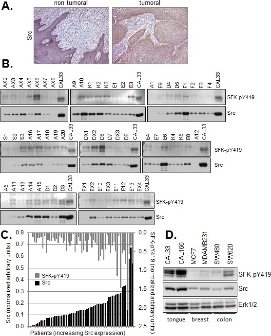
(A) Immunohistochemical staining of Src in a representative formalin fixed paraffin embedded (FFPE) human Squmaous Cell Carcinoma of the tongue. (bar=200μm) (B) Western blot analysis of Src and phospho-SFK (SFK-pY419) in membrane preparations of human head and neck tumor samples (10μg). Controls correspond to membranes (10μg) prepared from exponentially growing CAL33 cells treated for 5 minutes with 30 ng/ml EGF. (C) Quantitative Western blot data were normalized to control values within each series of 6-8 patient samples and plotted with respect to Src expression. Inter-assay variability calculated on control values from six independent runs was <27%. Bars represent single patient samples. (D) Western blot of Src and phospho-SFK (SFK-pY419) in total cell lysates of the indicated human tumor lines.