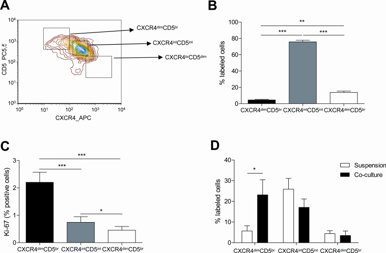
Figure 3. The proliferative CXCR4dimCD5br compartment of CLL cells is promoted by the co-culture with BMSC, CD40L and CpG ODN.
PBMC from 40 patients diagnosed with CLL were used to analyze the expression of CXCR4 and CD5 by FC. (A) Representative contour plot of CXCR4 and CD5 expression by CLL cells from one patient. (B) Mean percentage ± SEM of CLL cells in the three compartments defined by CXCR4 and CD5 densities are depicted in the graph (**P<0.01, ***P<0.001, one-way ANOVA). (C) Mean percentage ± SEM of Ki-67 expression in the three compartments (*P<0.05, **P<0.01, ***P<0.001, one-way ANOVA). (C) Primary CLL cells from 7 patients cultured in suspension or in co-culture with BMSC, CD40L and CpG ODN for 48 hours were used to analyze the expression of CXCR4 and CD5. (*P<0.05, two-way ANOVA, Bonferroni's post-test. Graph shows mean ± SEM).

