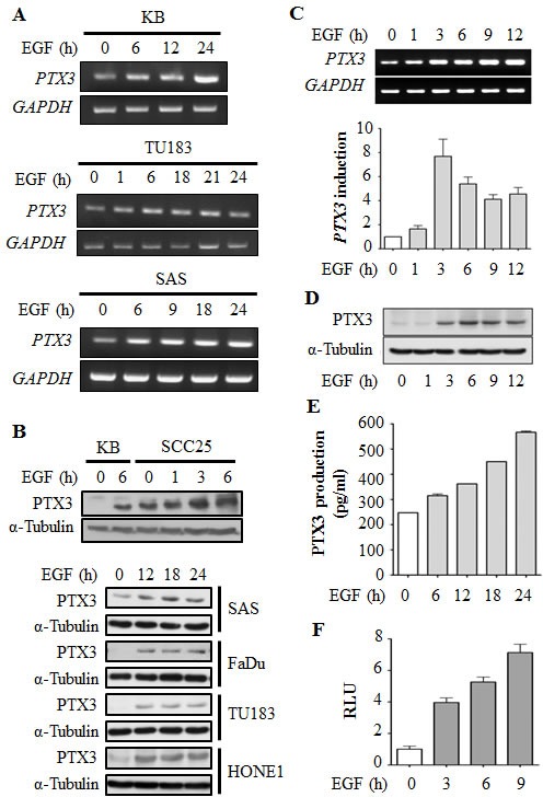
Figure 1. EGF induces transcriptional activation of PTX3 gene expression in head and neck squamous cell carcinoma (HNSCC) cell lines.

(A) HNSCC cell lines were treated with 50 ng/ml EGF for a period of time as indicated. Expressions of PTX3 and GAPDH mRNA were analyzed by an RT-PCR and examination in 2% agarose gels. (B) Lysates of cells were prepared and subjected to SDS-PAGE and analyzed by Western blotting with antibodies against PTX3 and α-tubulin. (C) KB cells were treated with 50 ng/ml EGF for a period of time as indicated. Expressions of PTX3 and GAPDH mRNA were analyzed by an RT-PCR (upper panel) and a real-time quantitative PCR (lower panel). Relative levels of PTX3 were normalized to GADPH. Values represent the mean ± S.E. of three independent experiments. (D) Lysates of EGF-treated KB cells were prepared and subjected to SDS-PAGE and analyzed by Western blotting with antibodies against PTX3 and α-tubulin. (E) KB cells were treated with 50 ng/ml EGF for a period of time, and then conditioned medium was collected to analyze PTX3 protein by an ELISA. Values represent the mean ± S.E. of three independent experiments. (F) KB cells were transfected with 0.5 μg PTX3 promoter construct by lipofection and then treated with 50 ng/ml EGF for various times as indicated. Luciferase activities and protein concentrations were then determined and normalized. Values represent the mean ± S.E. of three determinations.
