
Figure 2. Activation of NF-κB is essential for EGF-induced PTX3 expression.
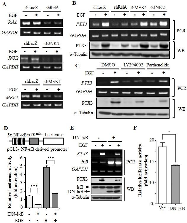
(A) RelA-, MEK1-, and JNK2-deficient cell lines were selected by infecting KB cells with a lentivirus containing an expression vector encoding short hairpin (sh)RNA against RelA (shRelA), MEK1 (shMEK1), and JNK2 (shJNK2). Expressions of RelA, MEK1, JNK2, and GAPDH mRNAs were analyzed by an RT-PCR and examined in 2% agarose gels. shLacZ, negative control. (B) shRNA containing cells was treated with 50 ng/ml EGF for 3 h, and expressions of PTX3 mRNA and protein were respectively analyzed by an RT-PCR and Western blotting (WB). shLacZ, negative control. (C) KB cells were treated with 25 μM LY294002, 10 μM parthenolide, or 0.1% DMSO for 1 h, followed by treatment with 50 ng/ml EGF for 3 h. Expressions of PTX3 mRNA and protein were respectively analyzed by an RT-PCR and WB. (D) The construct of the pTK promoter with five repeated NF-κB-binding sites bearing the luciferase gene is presented (upper panel). KB cells were transfected with 0.5 μg pTK-NF-κB promoter, 1 μg dominant negative IκB (DN-IκB) expression vector, and 1 μg control vector by lipofection and then treated with 50 ng/ml EGF for 6 h. Luciferase activities and protein concentrations were then determined and normalized (lower panel). (E) KB cells were transfected with 1 μg DN-IκB expression vector or 1 μg control vector by lipofection and then treated with 50 ng/ml EGF for 6 h before extraction of RNA or lysates. Expressions of PTX3, IκB, GAPDH, and α-tubulin mRNAs and proteins were respectively analyzed by an RT-PCR (PCR) and Western blotting (WB). (F) KB cells were transfected with 0.5 μg PTX3 promoter construct, 1 μg DN-IκB expression vector, or 1 μg control vector by lipofection and then treated with 50 ng/ml EGF for 6 h. Luciferase activities and protein concentrations were then determined and normalized. Values represent the mean ± S.E. of three determinations.