
Figure 3. The binding of c-Jun to AP1-binding site located on the PTX3 promoter is essential for EGF-induced promoter activity.
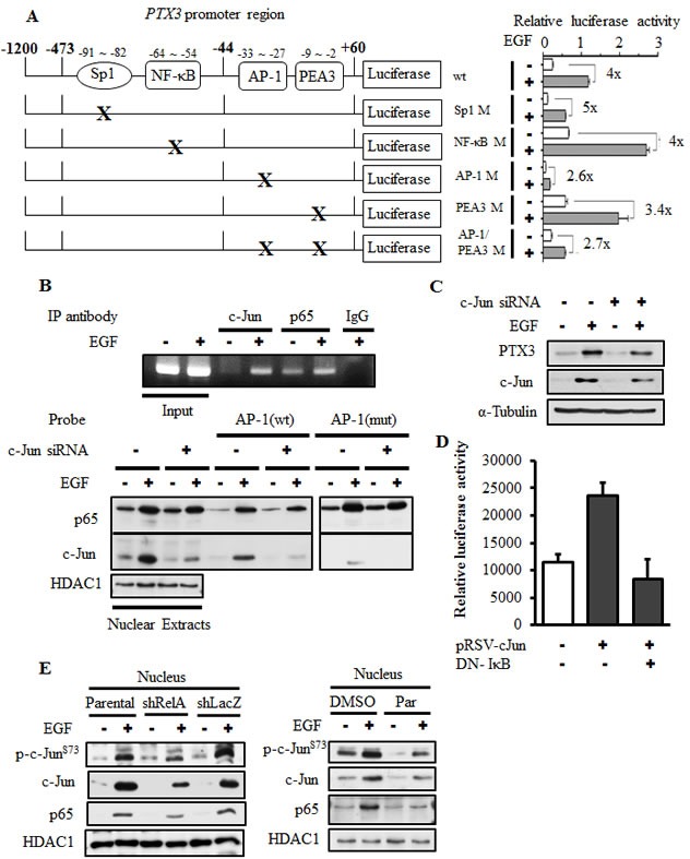
(A) Schematic representation of reporter constructs with mutations of transcriptional factor-binding sites shown on the left. Luciferase activities of these reporter constructs in KB cells treated with 50 ng/ml EGF for 6 h are shown on the right. Values of luciferase activities are presented as the mean ± S.E. of three determinations. (B) KB cells were transfected with or without 20 nM c-Jun siRNA oligonucleotides by lipofection. After 50 ng/ml EGF treatment for 1 h, nuclear extracts were prepared, and chromatin immunoprecipitation (ChIP) (upper panel) and DNA affinity precipitation (lower panel) assays were performed as described in “Materials and methods”. Binding of p65 and c-Jun to the AP1 site of PTX3 promoter region (AP1 (wt)) and mutant probe (AP1 (mut)) was analyzed by Western blotting. HDAC1, loading control. (C) KB cells were transfected with 20 nM c-Jun siRNA oligonucleotides by lipofection. After 50 ng/ml EGF treatment for 6 h, expressions of PTX3, c-Jun, and α-tubulin were analyzed by Western blotting (WB). (D) KB cells were transfected with 0.5 μg PTX3 promoter construct, 1 μg DN-IκB expression vector, and 1 μg pRSV-c-Jun by lipofection. Luciferase activities and protein concentrations were then determined and normalized. Values are represented as the mean ± S.E. of three determinations. (E) Parental (wt) and shRelA cells were treated with 50 ng/ml EGF for 6 h. Expressions of p65, c-Jun and phosphorylation of c-Jun on Ser73 were analyzed by WB. shLacZ, negative control. HDAC1, loading control.