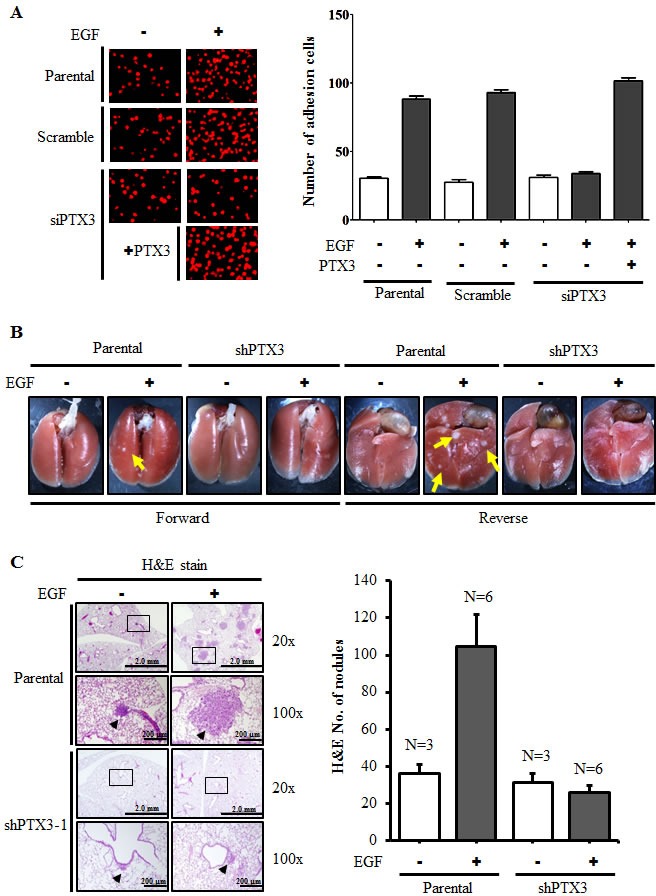
Figure 5. PTX3 mediates EGF priming for tumor dissemination to the lungs.

(A) FaDu cells were transfected with 30 nM PTX3 siRNA oligonucleotides by lipofection. After 50 ng/ml EGF and 250 ng/ml PTX3 treatment for 3 h, cells were then labeled with DiI and cultured with endothelial cells for 3 h. The attachment of cells was examined using a microscope (left panel). The number of attached cells was counted using three randomly chosen fields under the microscope from three independent experiments (right panel). Values represent the mean ± S.E. of three determinations. (B) A lung-colonization analysis was performed by injecting 2 × 10⁵ FaDu cells into a lateral tail vein of mice. Prior to the injection, cells were treated as indicated with 50 ng/ml EGF for 3 h. Nodules were examined and photographed at 2 month. Arrows point to metastatic nodules. (C) The images of tumors (left panel) and numbers of nodules (right panel) were examined using H&E staining and counted under a microscope, respectively. Error bars indicate SEM. N, number of SCID mice.
