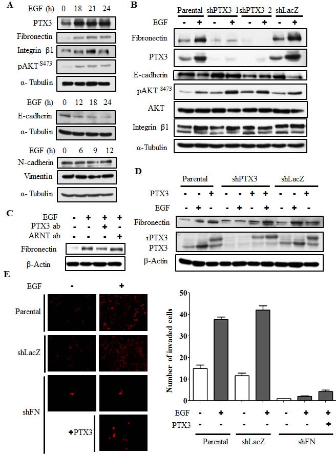
Figure 6. EGF-induced PTX3 regulates expressions of fibronectin and E-cadherin.

(A) HONE1 cells were treated with 50 ng/ml EGF for a period of time as indicated. Lysates of cells were prepared and subjected to SDS-PAGE and analyzed by Western blotting with antibodies against PTX3, fibronectin, integrin β1, phosphorylation of Akt at serine 473, E-cadherin, N-cadherin, vimentin, and α-tubulin. (B) Parental and shPTX3 HONE1 cells were treated with 50 ng/ml EGF. Lysates of cells were prepared and subjected to SDS-PAGE and analyzed by Western blotting with antibodies against PTX3, fibronectin, phosphorylation of Akt at serine 473, E-cadherin, integrin β1, and α-tubulin. shLacZ, negative control. (C) HONE1 cells were treated with 50 ng/ml EGF and 15 μg/ml anti-PTX3 antibodies or 15 μg/ml aryl hydrocarbon receptor nuclear translocator (ARNT) antibodies as negative control. Lysates of cells were prepared and subjected to SDS-PAGE and analyzed by Western blotting with antibodies against fibronectin and β-actin. (D) Parental and shPTX3 HONE1 cells were treated with 50 ng/ml EGF and 250 ng/ml PTX3 protein. Lysates of cells were prepared and subjected to SDS-PAGE and analyzed by Western blotting with antibodies against PTX3, fibronectin and β-actin. shLacZ, negative control. (E) Transendothelial invasion assay was performed as described in “Material and methods”. Parental and fibronectin knockdown (shFN) HONE1 cells were treated with 250 ng/ml PTX3 and 50 ng/ml EGF for 6 h and stained with DiI, and then were loaded in the upper chamber of filter inserts. After incubation for 48 h, the non-invasive cells were removed. The invasive images were examined using a microscope (left panel). Original magnification, ×200. The number of invasion cells was counted using three randomly chosen fields under the microscope from three independent experiments (right panel). Error bars indicate SEM. shLacZ, negative control.
