
Figure 7. EGF-induced PTX3 regulates the expression of MMP9.
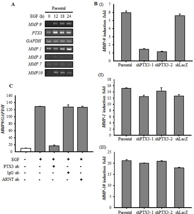
(A) HONE1 cells were treated with 50 ng/ml EGF for a period of time as indicated. Expressions of PTX3, MMP1, MMP3, MMP7, MMP9, MMP10 and GAPDH mRNA were analyzed by RT-PCR and examined in 2% agarose gel. (B) Parental and shPTX3 HONE1 cells were treated with 50 ng/ml EGF for 6 h. The expression of MMP9 (I), MMP1 (II) and MMP10 (III) mRNA was also analyzed by Real-time quantitative PCR. Relative levels of MMPs were normalized by GADPH. Error bars indicate SEM of three independent experiments. shLacZ, negative control. (C) NONE1 cells were treated with 15 μg/ml anti-PTX3 antibodies, 15 μg/ml immunoglobulin G (IgG), 15 μg/ml anti-aryl hydrocarbon receptor nuclear translocator (ARNT) antibodies and 50 ng/ml EGF for 9 h. The expression of MMP9 mRNA was also analyzed by Real-time quantitative PCR. Relative levels of MMP9 were normalized by GADPH. Error bars indicate SEM of three independent experiments.