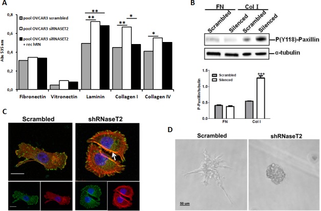
Figure 7. RNASET2 silencing affects both cell adhesion and FA dynamics in OVCAR3 cells.

A) A colorimetric in vitro cell-adhesion assay was performed on five different ECM components. Significant differences in cell-adhesion on laminin, collagen type I and collagen type IV were observed when RNASET2-silenced OVCAR3 cell clones were compared to control clones. Such differences turned out to be weakened when RNASET2-silenced cells were exposed to recombinant RNASET2 protein. Triplicate experiments were performed with four RNASET2-silenced and four control OVCAR3 clones. Statistical analysis was performed using two-tailed Student's t-test. * p<0,05; ** p<0,01. B) Evaluation of phoshoryated paxillin on total cell lysates upon adhesion on fibronectin (FN) or collagen type I (Col I). Upper panel: immunoblotting for the expression of phospho-paxillin. Lower panel: densitometric analysis reporting the levels of phosphorylated paxillin with respect to α-tubulin expression of cells grown on ECM matrices. Statistical analysis of data from triplicate experiments was performed using Student's t-test. *** p< 0.001. C) Confocal IIF performed on migrating OVCAR3 cell pools. Starved cells were induced to migrate through a wound upon stimulation with FCS for 24 hr, and then fixed with paraformaldehyde. FAs were stained with anti-phoshphorylated paxillin (green); actin was stained with phalloidin (red). Nuclei were counterstained with DAPI. The white arrow indicates dorsal stress fibers present in RNaseT2-silenced cells. Scal bar: 50 μm. D) Control vs. RNASET2-silenced OVCAR3 cells were grown in reduced growth factor-matrigel 3D culture and pictures were taken with a light microscope.
