
Figure 5. Modulation of LMWPTP results in impaired migration and invasion in colorectal cancer cells.
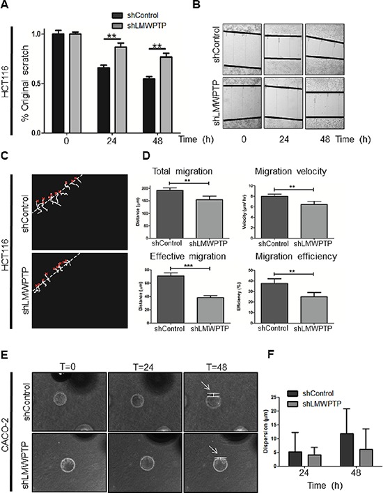
(A, B) HCT116 cell migration was measured by scratch assays, where simple scratch wounds were made using a pipet tip, and pictures are taken at 0 h, 24 h, and 48 h. Persistent area of clear plastic was measured and statistical analysis was performed using student's T-test. (C, D) Two-dimensional migration was analyzed using a ring-barrier system. HCT116 cell migration on gelatin was tracked during 24 h, with locations being captured using time-lapse microscopy every 12 min (x=start, line=cell track) (C). Quantification of migrated path indicates that the total migration and velocity were significantly reduced in LMWPTP knockdown cells. Effective migration and thereby efficiency are even further reduced. (D; *P < 0.05; **P < 0.01; ***P < 0.001). (E, F) Beads were coated with either CACO-2 LMWPTP knockdown or control cells for 24 hours, and embedded in a collagen gel matrix. Cells were allowed to invade the collagen matrix, and pictures were taking at 0 h, 24 h, and 48 h (examples in E). The cell dispersion from the bead (arrow) into the collagen matrix was measured, and a trend towards reduced invasion was observed in LMWPTP knockdown cells. Data represents at least four beads (F).