
Fig. 7.
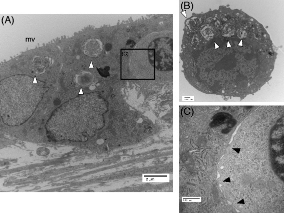
Transmission electron micrographic images of AECs in direct contact with BMSCs on day 14. a Type II-like epithelial cells observed in this preparation. b Freshly isolated type II AECs. The lamellar bodies that were observed in the freshly isolated cells were also observed in some of the epithelial cells in a (a, b white arrow heads). c A magnified image of the cellular contact observed in the black rectangle shown in a. The black arrow heads indicate contacts between the epithelial and mesenchymal cells. mv microvilli