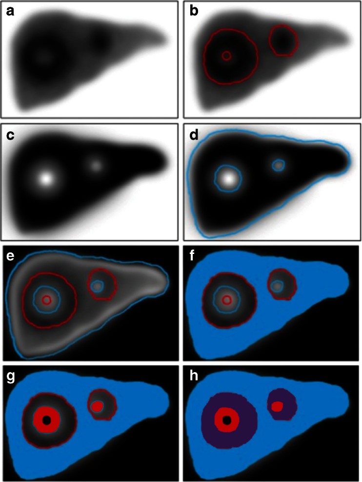
Fig. 1.
A threshold applied to the TcMAA SPECT image (a) defines the MAA-positive volume (b), and to the 99mTc-sulphur colloid (SC) SPECT (c) defines the SC-positive volume (d). Coregistration of the two SPECT scans results in four compartments: f unirradiated functional liver (V FL-UN), MAA-negative, SC-positive (blue), g tumour (V T), MAA-positive SC-negative (red), h irradiated functional liver (V FL-IR), MAA-positive SC-positive (purple), and tumour necrosis (V NULL), MAA-negative SC-negative (black)

