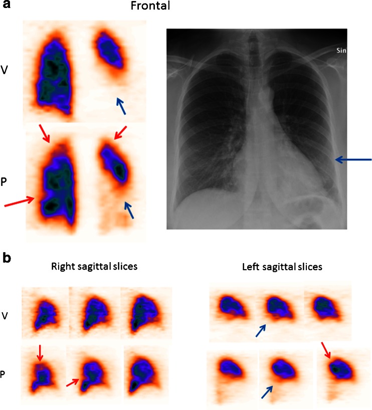
Fig. 1.
Frontal (a) and sagittal (b) slices from V/P SPECT and chest X-ray in a pregnant woman diagnosed with both PE and pneumonia in the second trimester of pregnancy. Red arrows indicate segmental perfusion defects in areas with preserved ventilation representing PE in the right and left lung. Blue arrows indicate absent ventilation and reduced perfusion representing pneumonia in the lower left lung

