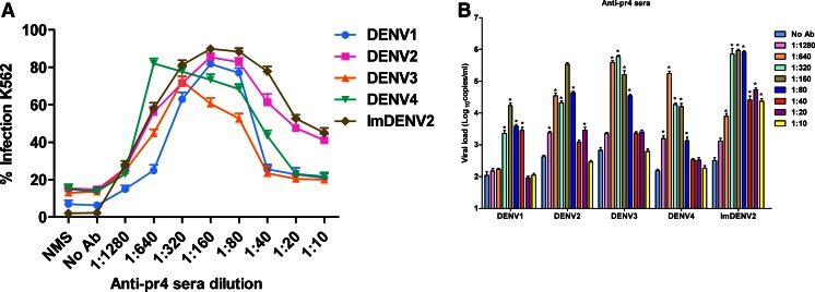
Fig. 7.
ADE of DENV infection in K562 cells mediated by anti-pr4 sera. Twofold serially diluted antibodies and an equal volume of DENV (MOI of 1) were mixed for 1 h at 37 °C and added to K562 cells. a The percent of infected K562 cells was measured at 3 days post-infection by flow cytometry. b Viral RNA levels of supernatants were accessed at 4 days post-infection by qRT-PCR. Anti-pr4 sera showed significant ADE activities toward standard DENV and imDENV2. NMS was used as control. Data are expressed as means of at least three independent experiments. The error bars represent standard deviations (SD). If there is no error bar, it is not that no variations among three independent experiments but that the variations are too small to show in the figure. *p < 0.05 vs No Ab

