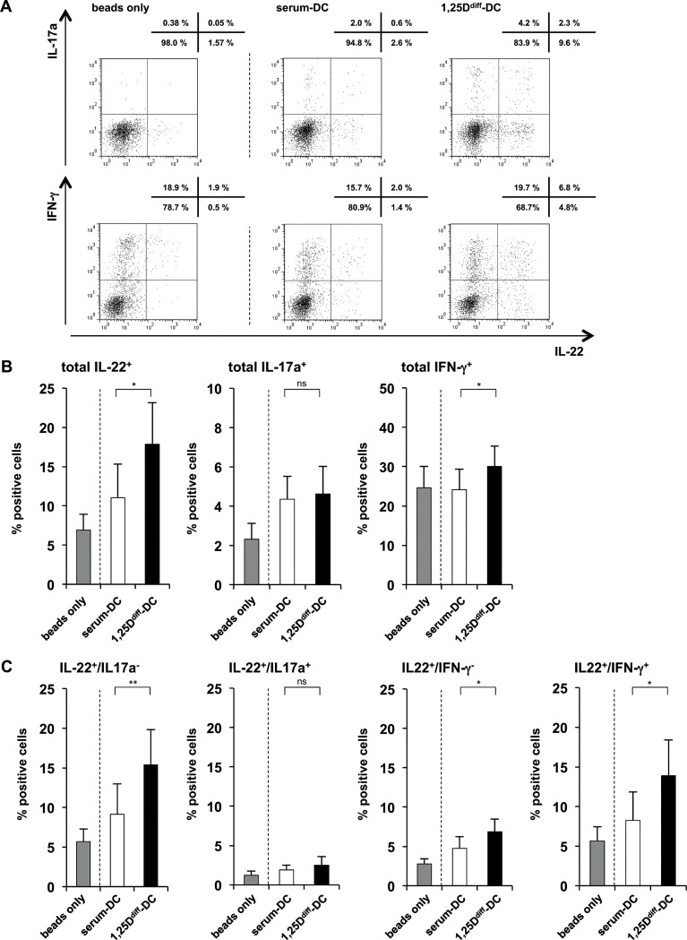
Fig 5. Supernatants of 1,25Ddiff-DCs promote differentiation of IL-22-expressing T cells.
Supernatants of TLR2/1-induced 1,25Ddiff-DCs and serum-DC were added to naïve CD4+ T cells activated with CD3/CD28-coated beads (as described in Fig 4). After five days, rIL2 was added to all cultures. On day 12, T cells were re-stimulated with PMA/Ionomycin for five hours, the last 2.5 hours of culture in the presence of Brefeldin A, in fresh media and intracellular cytokine expression of IL-22, IFN-γ or IL-17a was measured. (A) Dot plots from one representative staining of one donor out of eleven. Upper panel of dot plots shows co-expression of IL-17a and IL-22, lower panel shows co-expression of IFN-γ and IL-22. Numbers above each dot plot indicate frequency of positive cells in each quadrant. (B) Frequency of total IL-22-, IL-17a- and IFN-γ-expressing CD4+ T cells assessed by intracellular cytokine staining (mean percentage of positive cells ± SEM, n = 11). (C) Frequency of IL-22+/IL-17a+ and IL-22+/IL-17a- or IL-22+/IFN-γ+ and IL-22+/IFN-γ- CD4+ T cells assessed by intracellular cytokine staining (mean percentage of positive cells ± SEM, n = 11). *p<0.05, **p<0.01

