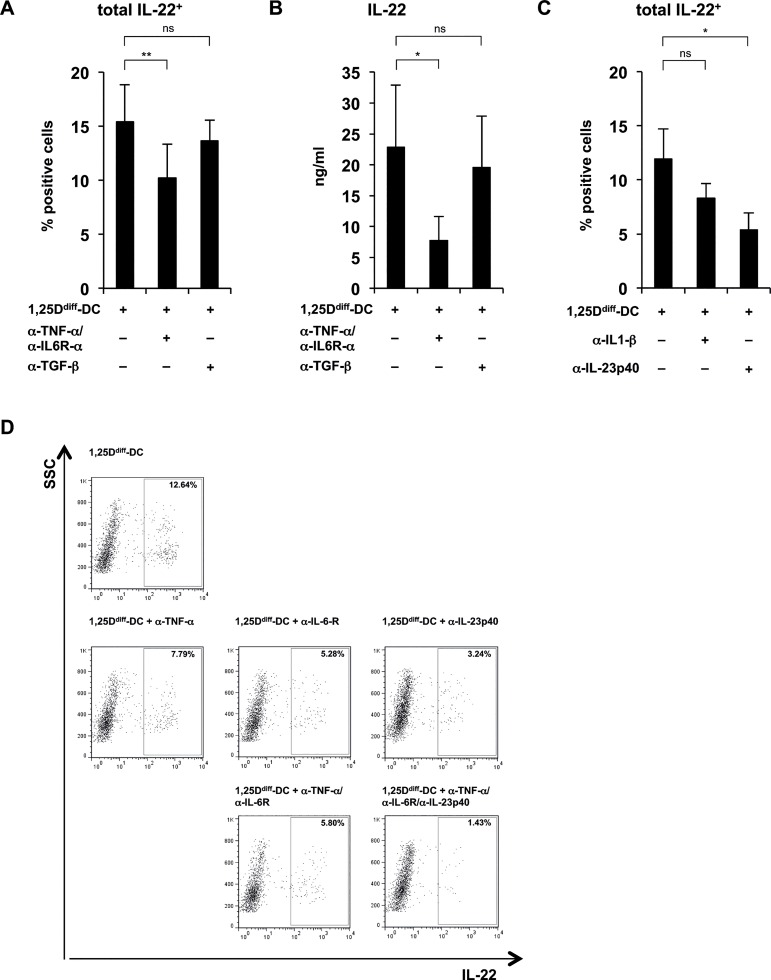
Fig 6. 1,25Ddiff-DC-supernatant mediated priming of IL-22-producing T cells is dependent on TNF-α IL-6 and IL-23.
Supernatants of TLR2/1-stimulated 1,25Ddiff-DCs were added to naïve CD4+ T cells activated via CD3/CD28-coated beads (as described in Fig 4) in the presence or absence of different monoclonal blocking antibodies as indicated. After five days, rIL2 was added to all cultures. On day 12, T cells were restimulated with PMA/Ionomycin for five hours, the last 2.5 hours of culture in the presence of Brefeldin A, in fresh media and intracellular cytokine expression of IL-22, IFN-γ or IL-17a was measured. Cytokine secretion was evaluated after 18–24 hours without further addition of Brefeldin A. (A) Anti-TNF-α, anti-IL-6R-α (5 μg/ml each) or anti-TGF-β (10 μg/ml). T cell-derived IL-22 assessed by ELISA (mean of cytokine levels in ng/ml ± SEM, n = 5). (B) Anti-TNF-α, anti-IL-6R-α (5 μg/ml each) or anti-TGF-β (10 μg/ml). Frequency of total IL-22-expressing CD4+ T cells assessed by intracellular cytokine staining (mean percentage of positive cells ± SEM, n = 5). (C) Anti-IL-1β or anti-IL-23p40 (5 μg/ml each). Frequency of total IL-22-expressing CD4+ T cells assessed by intracellular cytokine staining (mean percentage of positive cells ± SEM, n = 5). (D) Anti-TNF-α, anti-IL-6R-α or anti-IL-23p40 (5 μg/ml each) blocking antibodies alone or in combination. Dot plots from one representative staining of one donor out of five showing the frequency of IL-22-expressing CD4+ T cells against the sideward-scatter (SSC). Numbers in rectangle gate indicate frequency of positive cells. *p<0.05, **p<0.01

