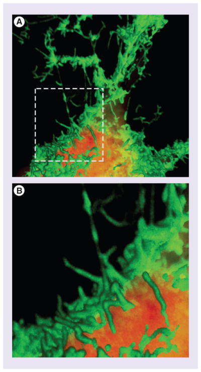
Figure 1. Expression of Ebola virus VP40 results in VLP formation and egress.

A GFP-VP40 fusion protein expressed inHEK293T cells shows robust virus-like particle assembly and egress at the plasma membrane (A). (B) corresponds to inset indicated in (A). Upper panel is approximately 21 μm wide. The cytoplasm is stained with HCS CellMask™ Deep Red. Images were rendered using Volocity software. This image was generated by the Freedman laboratory in the PennVet Imaging Core Facility.
