
Fig 2. Three-dimensional structure of the PvDBPII dimer.
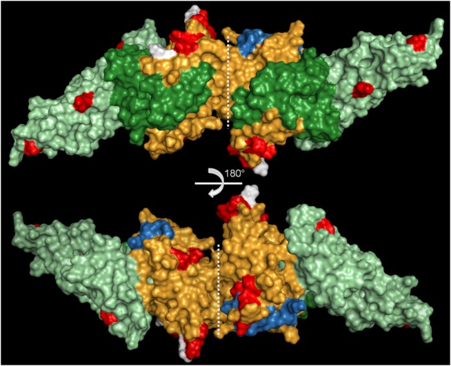
PvDBPII dimer (3RRC) showing the polymorphic sites in P. vivax (red) and P. simium (white). The three subdomains of the protein are shown in green (subdomain 1), orange (subdomain 2) and light green (subdomain 3). Residues important for the DARC interaction are shown in blue. 3-D structure visualized using PyMol. The structures correspond to a 180° rotation in the horizontal plan.