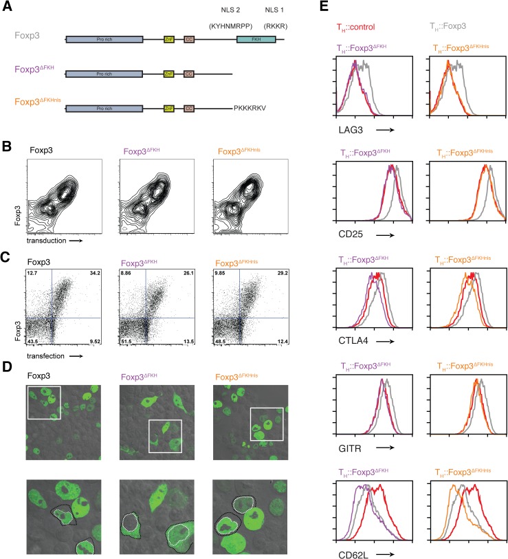
Fig 2. The role of nuclear localisation signals in the forkhead domain of Foxp3.
(A) Schematic illustrations of nuclear localization signals and addition of SV40 NLS. (B) Expression of Foxp3, Foxp3ΔFKH and Foxp3ΔFKHnls and the rCD8 reporter in transduced primary T cells. (C) Expression of Foxp3, Foxp3ΔFKH and Foxp3ΔFKHnls and the rCD8 reporter in transfected HEK293 cells. (D) Subcellular localisation of the Foxp3, Foxp3ΔFKH and Foxp3ΔFKHnls stained with alexa488 anti-Foxp3 antibody in transfected HEK293 cells analysed by confocal microscope. Lower panel is a higher magnification of the boxed region in the upper panels with some cells having their nuclei outline with a dotted white line and the cell boundary with a dotted black line. (E) The effect of presence or absence of nls on expression of key molecules on the expression of key Foxp3 target genes. The expression of LAG3, CD25, CTLA4, GITR and CD62L was analysed by flow cytometry 48 h post-transduction in rested TH::Foxp3, TH::Foxp3ΔFKH and TH::Foxp3ΔFKHnls cells. Plots were gated on transduced cells that were rCD8a+ and are representative of three independent experiments.

