Figure 6.

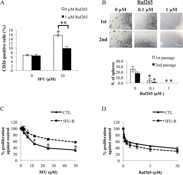
Anti-proliferative effect of Raf265 on CD26+ cells. A. Cells were treated with different combinations of Raf265 and 5FU. The percentage of CD26+ cells was determined. B. CD26+ cells with single cell suspensions were treated with different concentrations of Raf265 for the sphere formation assay. After 14 days, spheres were disaggregated and reseeded for the second passage of sphere formation. Representing images under a phase-contrast microscopy at 40× magnification were shown at the upper panel. The numbers of spheres formed were then quantified after 14 days of incubation and the bar chart presenting the numbers of spheres formed was shown at the lower panel. HT29 cells with 5FU resistance were treated with C. 5FU at 0–50 μM and D. Raf265 at 0–10 μM and MTT assay was performed. Data are presented as means ± SD from three independent experiments and statistical analysis was performed by one-way ANOVA. *p < 0.05 versus untreated control.
