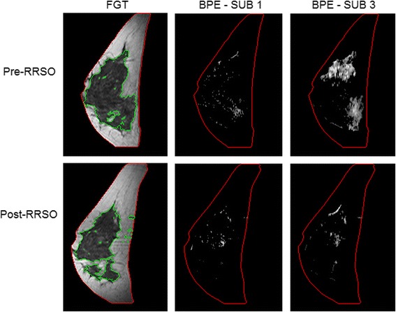
Fig. 4.

Representative examples of fibroglandular tissue (FGT) and background parenchymal enhancement (BPE) from a magnetic resonance imaging (MRI) slice obtained pre-RRSO and post-RRSO in a woman who did not develop breast cancer after undergoing risk-reducing salpingo-oophorectomy (RRSO). FGT is circumscribed by green contours. BPE is estimated in both the first and third subtraction series (SUB 1 and SUB 3, respectively). This 40-year-old (at time of RRSO) woman had her pre-RRSO MRI at 6 months before RRSO and her post-RRSO MRI at 1 month after RRSO. She had no personal history of breast cancer or other cancer and had no breast cancer diagnosis for up to 9 years of post-RRSO follow-up. This example illustrates that there was a decrease of BPE and FGT after her RRSO. The volumetric pre-RRSO BPE % values were 1.5 % (SUB 1) and 6.5 % (SUB 3) and after RRSO, and BPE % values were 0.6 % (SUB 1) and 3.6 % (SUB 3)
