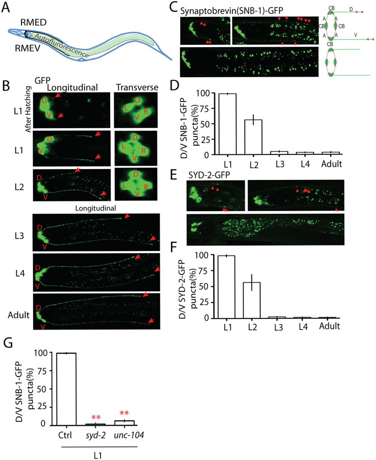
Figure 1. RMED and RMEV neurons form transient clusters of presynaptic components at dendritic D/V neurites during larval development.

(A) Illustration of the morphology of RME neurons. RME neurons (black color) locate in the head region of C. elegans, and RME axons form a ring-shape bundle at the nerve ring region. RMED and RMEV grow two posterior neurites along the dorsal/ventral cords. When imaging RME neurons, the gut autofluorescence is often seen between the dorsal and ventral cords (B, C and E.). (B) The development of RME D/V neurites visualized by a marker expressing GFP in RME neurons (Punc-25∷GFP). The four RME neurons migrated to their final position before hatching, but the development of D/V neurites occurred after hatching. Red arrows label the end of D/V neurites. D: RMED, V: RMEV, L: RMEL, R: RMER. (C-F) RME D/V neurons form transient synapses at early developmental stages. At the first larval (L1) stage, synaptic vesicles and synaptic active zones are clustered at dorsal/ventral neurites. No synaptic vesicle and synaptic active zone puncta were observed in L3 worms. Representative images and schematics show the localization of synaptic vesicle puncta (Synaptobrevin (SNB-1)-GFP, C) and synaptic active zone puncta (SYD-2-GFP, E) in RME neurons. CB, cell bodies; A, axons; D/V, dorsal/ventral neurites; red dot, synaptic puncta. Red arrowheads highlight the transient clusters of presynaptic components. (D and F) Quantification of the percentage of animals with D/V puncta. (G) Quantification data show that loss-of-function in syd-2 and unc-104 causes absence of SNB-1-GFP puncta in D/V neurites of L1 animals. Experiments were performed at least 3 times, with N ≥80 animals each time. Data is shown as mean ± SD. Student test, ** P < .01. Scale bar, 10 μm.
