Fig. 1.

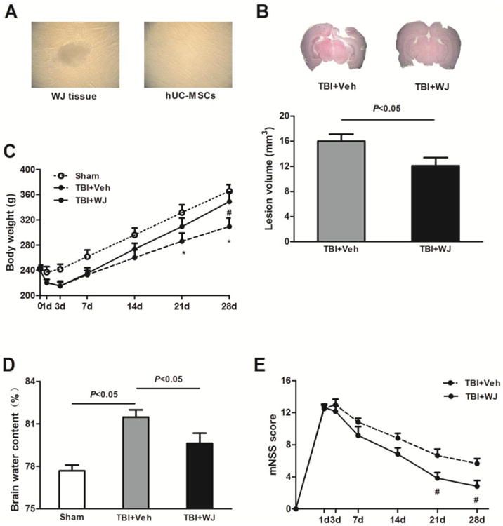
Wharton's jelly (WJ) tissue transplantation decreases lesion volume and brain edema and improves neurologic deficits. (A) Single-spindled or triangular hUC-MSCs adhered rapidly around the WJ tissue and proliferated rapidly without visible changes in growth pattern or morphology. (B) Top: Representative brain sections from WJ tissue-treated and vehicle-treated rats on day 28 after traumatic brain injury (TBI). Bottom: Lesion volume was smaller in WJ tissue-treated rats than in vehicle-treated rats (n=6 rats per group, t-test). (C) The body weight of WJ tissue-treated TBI rats recovered faster and increased more than that of vehicle-treated rats during the 28 days after TBI (n=6 rats/group, *p<0.05 vs. sham group, #p<0.05 vs. vehicle-treated TBI group, two-way ANOVA followed by Bonferroni post-hoc tests). (D) Brain water content was greater in the vehicle-treated TBI group than in the sham group but was reduced by WJ tissue treatment.n=6 rats/group, one-way ANOVA followed by Newman-Keulstest. (E) WJ tissue-treated rats had significantly less neurologic deficit than did vehicle-treated rats on days 21 and 28 after TBI. n=6 rats/groups, #p<0.05, two-way ANOVA followed by Bonferroni post-hoc test. Values are mean ± SD, mNSS, modified neurologic severity score.
