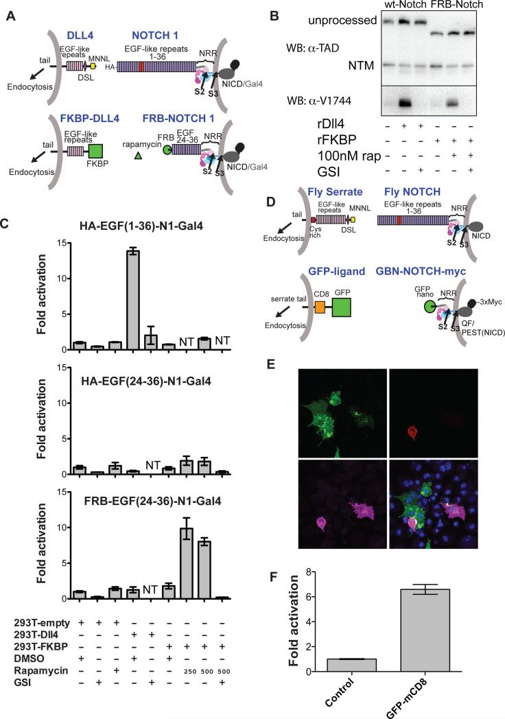
Figure 3. Development and evaluation of two synthetic Notch signaling systems.
(A) Schematic comparing the natural human Notch-ligand signaling system (top; EGF repeats 11-12 in red) to a synthetic signaling system placing NRR proteolysis under rapamycin-inducible control (bottom). Here, FKBP replaces the N-terminal portion of DLL4, and FRB replaces EGF-like repeats 1-23 of Notch1. The Notch1 ankyrin domain is also replaced with Gal4, as above (Malecki et al., 2006). (B) Western blots monitoring receptor proteolysis. U2OS cells stably expressing wild-type or FRB-Notch1 were grown in the presence of the DLL4 ectodomain or FKBP immobilized on plastic tissue culture dishes in the absence or presence of rapamycin (250 nM) and/or a GSI (Compound E, 400 nM). Blots were probed with an antibody directed against an epitope of intracellular Notch1 (α-TAD), or the α-V1744 antibody to S3-cleaved Notch1 (Cell Signaling). (C) Cell-based reporter gene assay. U2OS cells stably transfected with the indicated Notch variants were co-cultured with 293T cells transiently transfected with the indicated ligands. Luciferase activity for each U2OS line is reported relative to co-culture with 293T cells transfected with empty vector. Error bars reflect the standard error of readings performed in triplicate. Additional control experiments are provided in Figure S3. (D) Schematic illustrating design of a GFP - GFP-binding nanobody (GBN) synthetic ligand-receptor pair. Full-length fly Serrate and Notch are shown for reference. The artificial ligand consists of GFP, CD8 and the Serrate-derived tail. The ectodomain of the Notch-derived molecule consists of the GFP binding nanobody (GBN) and the NRR, and the intracellular domain contains the QF transcription factor, the Notch PEST domain, and a triple Myc tag. (E) Co-culture assay. S2R+ cells expressing GFP-mcd8-Ser as ligand (green, upper left panel) were co-cultured with cells expressing GBN-FlyNotch(NRR)-QF-3XMyc (GBN-N-QFMyc). Receptor is stained with anti-myc antibody (magenta, lower left panel). The tdTomato reporter signal is red (upper right panel). DNA was stained with DAPI (blue, lower right panel). (F) QAS luciferase readout of cell-mixing experiment. Luciferase reporter gene activity for the GBN-Notch cell line is reported relative to co-culture with control cells. Error bars represent the standard error of measurements performed in quadruplicate. Additional supporting data are provided in Figure S3.

