FIGURE 2.

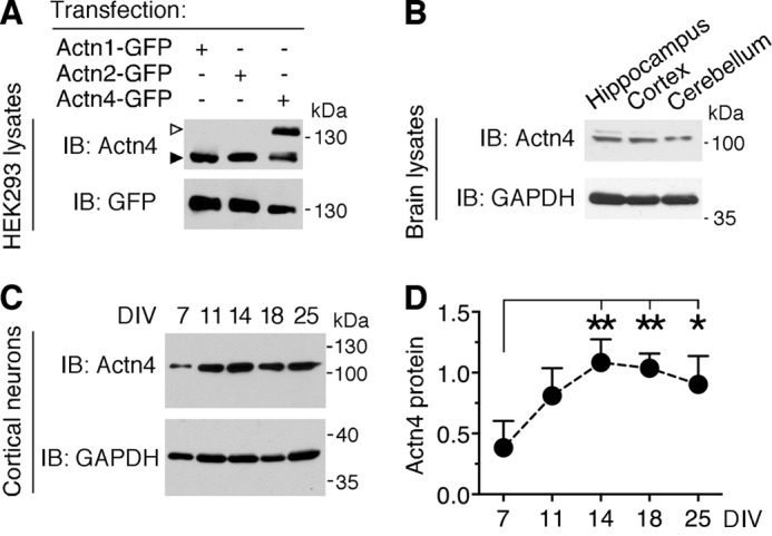
Actn4 is broadly expressed in the brain and in primary neurons at different stages of maturation. A, specificity of anti-Actn4 antibody. Shown are representative immunoblots (IB) of lysates from HEK293 cells transfected with Actn1-GFP, Actn2-GFP, or Actn4-GFP probed with anti-Actn4 and anti-GFP antibodies. Empty arrowhead, Actn4-GFP; black arrowhead, endogenous Actn4 present in HEK293 cells. B, representative immunoblots of lysates probed with anti-Actn4 and anti-GAPDH demonstrating Actn4 expression in adult mouse hippocampus, cortex, and cerebellum. C, developmental expression of Actn4 in primary cortical neurons. Shown are representative immunoblots of lysates from neurons at the indicated DIV probed with anti-Actn4 and anti-GAPDH. D, quantification of Actn4 expression relative to the loading control in cortical neurons at different ages in vitro. Mean ± S.E.; n = 3 independent experiments; *, p < 0.05, **, p < 0.01; one way analysis of variance.
