FIGURE 8.
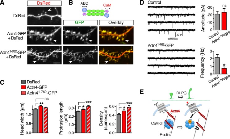
Actn4 regulates spine morphogenesis. A, representative Z-projections of confocal image stacks displaying dendritic segments from cortical neurons transfected with the indicated plasmids. Scale bar = 5 μm. B, schematic of native Actn4 homodimers. ABD, actin-binding domain. C, quantification from images like those in A of spine head width (left panel), protrusion length (center panel), and density (right panel). DsRed, n = 29 cells; Actn4-GFP, n = 23; Actn41-762-GFP, n = 21; *, p < 0.05; **, p < 0.01; ***, p < 0.001; ns, not significant. D, representative traces (left panel) and summary data (right panel) showing a reduction in the frequency of mEPSC recorded from cortical neurons transfected with Actn41-762-GFP compared with control neurons transfected with GFP (p = 0.00728, n = 10 cells/group). Calibration bar = 30 pA, 500 ms. E, working model of potential mechanisms underlying group 1 mGluR-dependent spine remodeling via the Actn4/CaMKII/F-actin interaction network. Actn4 enables the maturation of dendritic protrusions in conjunction with CaMKII by supporting CaMKII recruitment to F-actin. Activation of group 1 mGluR uncouples CaMKII from Actn4, potentially releasing it from F-actin, thereby enhancing spine dynamics by enabling remodeling of the actin cytoskeleton.

