FIGURE 8.
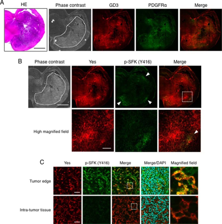
Yes was activated at the invasion front of glioma tissues. A, a coronal section of PDGFB-transfected gliomas generated in a p53−/− Gtv-a mouse at 3 weeks after the transfection was stained with an anti-GD3 mAb (red) and an anti-PDGFRα antibody (green). The closed dashed area represents a location of the tumors in a phase contrast image. Scale bars, 2 mm. B, sequential sections of PDGFB-transfected glioma tissues as shown in A. Double-immunostaining of Yes (red) and phosphorylated-Src family kinases (p-SFKs) (green) was performed. High-magnified vision fields of the dashed square region as indicated in Merge were presented in the lower panels. The arrowheads indicate activated Yes staining at the tumor edge. Scale bars, 2 mm (upper panels) and 200 μm (lower panels). C, double-immunostaining of Yes (red) and phosphorylated-Src family kinase (p-SFKs) (green) at the tumor edge, and the intra-tumor tissue was shown. High-magnified fields of dashed square areas as indicated in Merge are shown (right). HE, hematoxylin-eosin. Scale bars, 40 μm.

