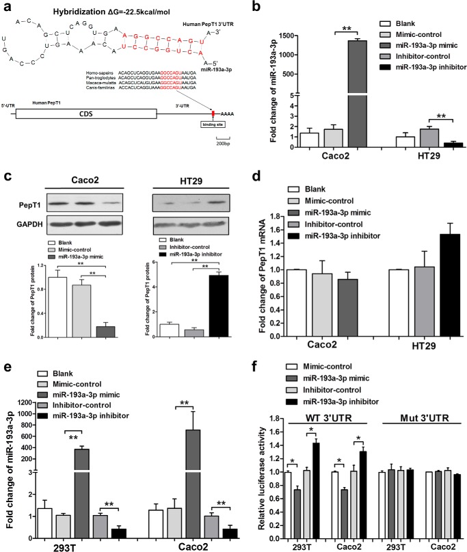
FIGURE 2.
Identification of PepT1 as a target of miR-193a-3p. A, schematic description of the conserved binding site for miR-193a-3p and human PepT1. B, qRT-PCR analysis of miR-193a-3p expression levels after transfection with the miR-193a-3p mimic, mimic-control in Caco2, the miR-193a-3p inhibitor, and inhibitor-control in HT29 cells normalized to U6. C, Western blotting of PepT1 protein expression levels after above transfection in Caco2 cells and HT29 cells. D, qRT-PCR analysis of PepT1 mRNA levels after above transfection in Caco2 and HT29 cells. E, qRT-PCR analysis of miR-193a-3p expression levels after transfection with the miR-193a-3p mimic, mimic-control, the miR-193a-3p inhibitor, and inhibitor-control in 293T and Caco2 cells. F, luciferase reporter activity after expression of the above transfections in 293T cells. Luciferase reporters carrying the wild-type (WT) or mutant (Mut) PepT1 3′-UTR were cotransfected into 293T and Caco2 cells along with the indicated oligonucleotides. *, p < 0.05; **, p < 0.01.

