
FIGURE 3.
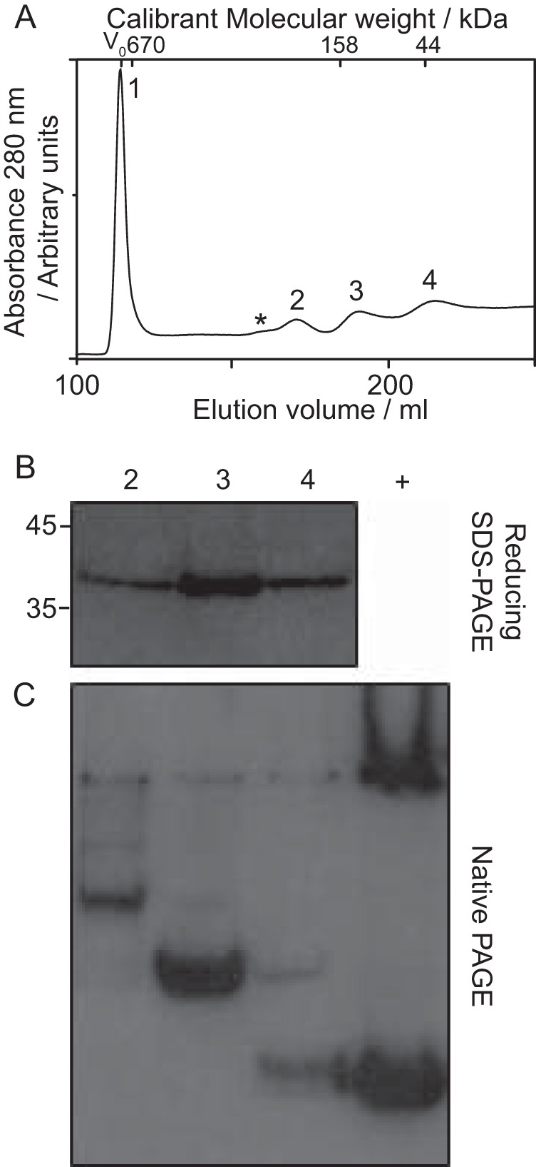
Disassembly of capsids composed of fused Cp. Capsids composed of fused dimers were disassembled with 4 m urea. A, size-exclusion chromatogram of fused Cp capsid disassembly reactions, resulting in a large peak consistent with non-dissociated capsid (Peak 1) and several other species labeled 2 to 4. Asterisk indicates additional low abundance species, which were not isolated. B, immunoblot of reducing SDS-PAGE of samples 2–4. All of the peaks contained protein of the same molecular weight, which was Cp antibody (10E11) reactive. Positions of standards are shown for reference. C, immunoblot of native PAGE of peak fractions. Proteins exhibited different migration patterns. The positive control (+) consists of Cp from disassembly of WT capsids.