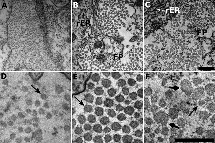
FIGURE 4.
BAPN precludes correct collagen fibril formation. Tendon construct cross-sections were investigated by TEM for their ultrastructure. A and D, 14-day control constructs show small diameter fibrils distributed in the extracellular space. At high resolution (D) the fibrils show almost circular outlines with large spacing in between individual fibrils (indicated by the arrow). B and E, at 21 days, the constructs show increased fibril diameters and tighter packing density. Moreover, the fibrils have similar fibril diameter with regular shapes. Rough endoplasmic reticulum (rER) indicate active cells, and fibripositors (FP) hint toward cell-collagen interactions and collagen production. A single collagen fibril is highlighted (arrow) that shows the typical close-to circular outline and is in regular distance to neighboring fibrils. C and F, BAPN-treated samples in contrast show heavily disrupted fibril shapes and irregular spacing in between individual fibrils. Fibril diameters vary considerably between very small (thin arrow) and very large fibrils (heavy arrows) of up to 100 nm. Moreover, the very large fibrils (heavy arrows) totally lost their circular shape. Compare the supplemental videos for a broader overview. Scale bar, 500 nm for A–C (in C); scale bar, 300 nm for (D–F) in F.

