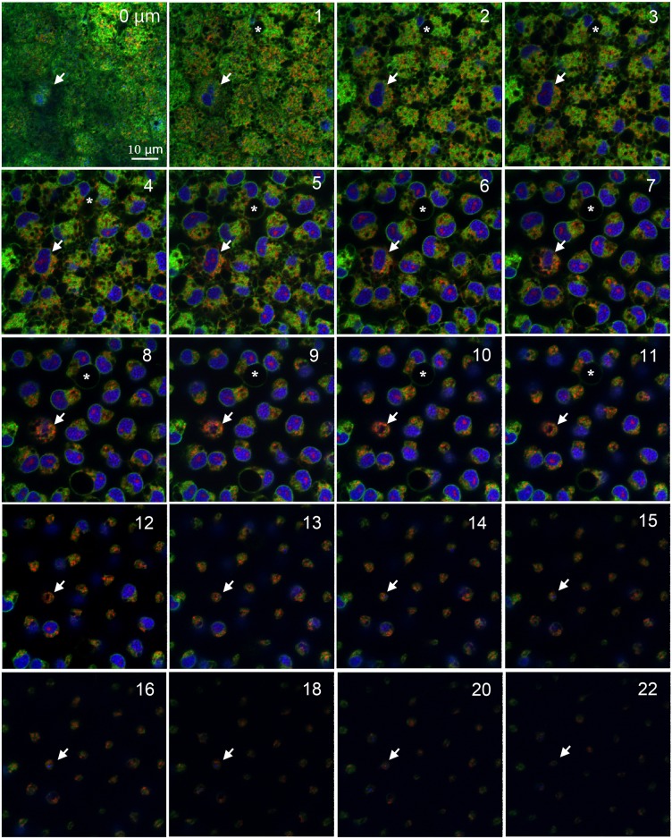
Fig 5. Deep serial cross-sections of pupal epithelial cells triple-stained with Hoechst 33342, BODIPY FL Thapsigargin, and MitoTracker Orange.
Numbers at the right upper corner indicate the depth from the apical cellular surface. White arrows indicate a large cell, likely a prospective scale cell. Asterisks indicate a large endosome-like or autophagosome-like unstained structure (see Fig 6). Red dots within a nucleus are not explainable. Also see S1 Movie.

