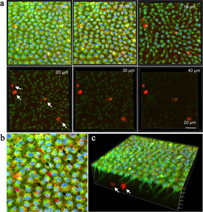
Fig 6. Deep serial cross-sections of pupal epithelial cells triple-stained with Hoechst 33342, BODIPY FL Thapsigargin, and LysoTracker Red.
A 3D image was reconstructed first, from which 2D cross-section images were presented in (a) and (b). (a) Serial images. Numbers at the right upper corner indicate the depth from the apical cellular surface. Small lysosomal bodies are strongly stained in red. Large endosome-like or autophagosome-like structures are weakly stained in red inside but their membranous structures are seen in orange. Hemocytes are strongly stained in red (arrows). (b) Enlarged image of (a), 5 μm in depth. Small lysosomal bodies are indicated by red arrows, whereas endosome-like or autophagosome-like structures are indicated by black arrows. (c) 3D image of an epithelial sheet. Stained hemocytes are indicated by arrows.

