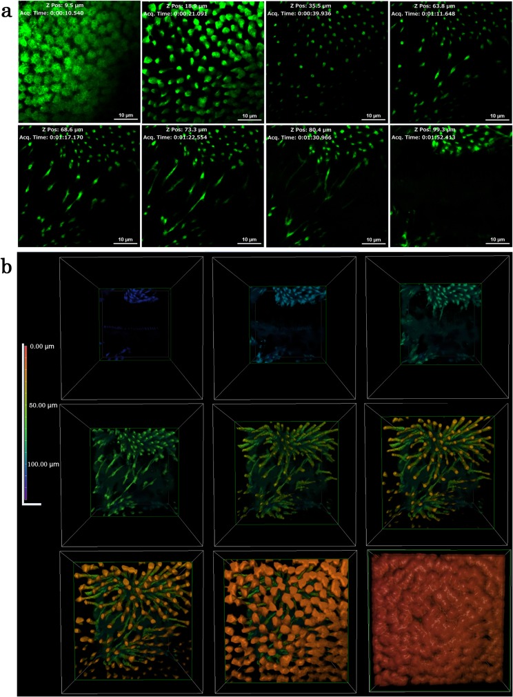
Fig 8. Deep serial cross-sections of pupal epithelial cells and a reconstruction of a stacking 3D image.
Cells were stained with CFSE for multiphoton microscopy. (a) Sections are presented from the top to the bottom. Also see S2 Movie. (b) Reconstruction of a stacking image from the bottom to the top. Also see S3 Movie.

