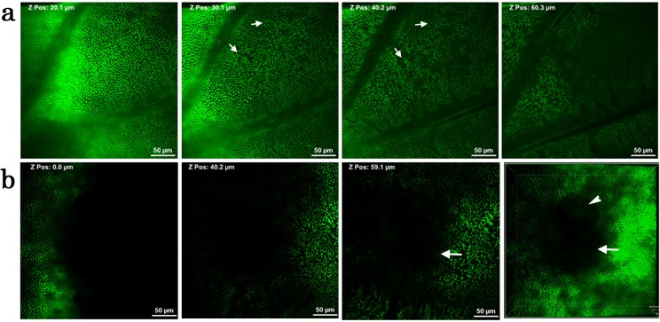
Fig 11. Wide-area images of deep sections of a pupal wing tissue.
Cells were stained with CFSE. (a) A basal wing area. Possible cellular clusters are indicated by white arrows. (b) A prospective eyespot area (white arrows). A bubble is trapped (an arrowhead). The rightmost panel is a synthetic image, showing a cone-like structure.

