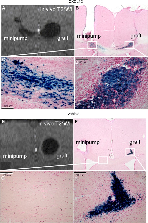
Fig. 5.

Corroboration of the chemotaxis by PB staining. a The NPC migration induced by CXCL12 infusion seen as the hypointensities on T2*WI. b The PB staining concurred with the hypointensities on T2*WI. c A magnified view of the migratory path toward the target. d A view of the graft. e T2*WI of the group with vehicle infusion showed little migration. f PB-stained cells were mainly distributed in the graft site. g A magnified view of the corpus callosum. h A view of the graft
