
Fig. 1.
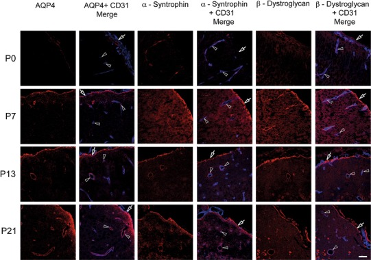
Members of the EBJC complex accumulate at brain surface and around vessels. Confocal images showing immunofluorescence labeling (red) of AQP4, α-syntrophin and β-dystroglycan in neocortex of postnatal mouse brain. These three members of the EBJC appear first at the brain surface and around penetrating vessels (lining the Virchow-Robins spaces), then around brain microvessels. Vessels (arrowheads) are identified by use of an endothelial marker (CD 31; blue). At P0, there was weak or no perivascular labeling for AQP4. At P7, AQP4 starts to appear around vessels (arrowheads), and at P13 and P21 there is AQP4 labeling around vessels of all calibers. Subpial AQP4 labeling (arrows) is present already at P0 and increases in strength towards P21. α-Syntrophin follows the same labeling pattern as AQP4. Weak labeling is observed for β-dystroglycan at P0. At P7, β-dystroglycan labeling is present at the brain surface (arrows) and around vessels of all calibers (arrow heads), thus being more extensive at this stage than the labeling for AQP4 and α-syntrophin. Scale bar 50 μm