
Fig. 5.
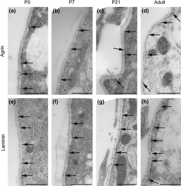
Agrin and laminin also occur in subpial basal lamina. Electron micrographs of immunogold labeling of agrin (a–d) and laminin (e–h). Both proteins are present in the basal lamina (arrows) opposed to subpial astrocyte endfeet. Labeling is distinct already at P0 and persists throughout the postnatal period. Scale bar 0.5 μm