
Fig. 1.
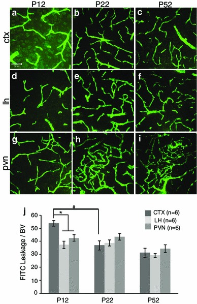
Postnatal blood–brain barrier development in the mouse cortex (CTX), lateral hypothalamus (LH) and paraventricular nucleus of the hypothalamus (PVN) at P12, P22 and P52. Example confocal images for each region are provided in panels a–i, and a quantitative summary by graph in j. There was a significant increase in extravascular FITC leakage in the CTX (a) compared to the LH (d) and PVN (g) at P12 (j; p < 0.05). Between P12 and P22 there was a significant decrease in extravascular FITC leakage specifically in the CTX (a, b; p < 0.05). At P22, there were no significant differences observed in extravascular FITC leakage between brain regions (b, e, h). At P52, there were no significant differences in extravascular FITC leakage (c, f, i) compared to P22 or between brain regions (j). Number of animals per group (n = 6) is provided in the code for the bars panel j. Significant differences between regions indicated by asterisk and for age as hash. Scale bar 50 µm in panel a, which applies to all images