
Fig. 6.
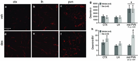
Prenatal exposure to dexamethasone (dex) impacted desmin-immunoreactive pericyte coverage in the mouse paraventricular nucleus of the hypothalamus (PVN) at P20. Example confocal images for each region are provided in panels a–f, and a quantitative summary by graph in g and h. In the PVN, there was a significant increase in desmin-immunoreactive pericyte coverage in dex-treated compared to vehicle-treated mice (c, f; *p < 0.01) when blood vessel density was taken into account (h; *p < 0.01). There were no significant differences observed in desmin-immunoreactive pericyte coverage in the cortex (CTX; a, d) or lateral hypothalamus (LH; b, e) between dex-treated or vehicle-treated mice. There was a significant increase in desmin-immunoreactive pericyte coverage in the PVN regardless of treatment compared to the CTX and LH (g). Number of animals per group is provided in the code for the bars in panels g and h. Significant differences between regions indicated by asterisk and for treatment as hash. Scale bar 50 µm in panel a, which applies to all images