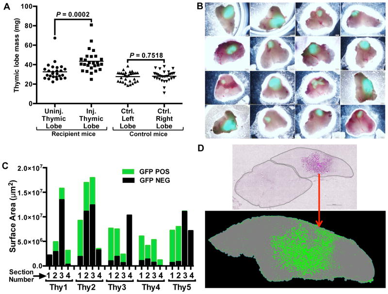
Figure 4.
Transplantation of fetal thymic cells drives growth of middle-aged thymuses. Single cell suspensions of thymic cells were prepared from GFP-transgenic E14.5 or E15.5 fetal thymuses and injected into one thymic lobe of each middle-aged (9–12 months old) WT female mouse. After 45 days, thymuses and spleens of recipient and control mice were removed and analyzed. (A) Injected (Inj.) and uninjected (Uninj.) thymic lobes, and lobes from control (Ctrl.) mice were separated and individually weighed. Each point represents the mass of an individual thymic lobe. Mean values for each group are shown as bars. (B) Representative recipient thymuses were imaged with an inverted fluorescence microscope 45 days after injection of dispersed GFP+ thymic cells. (C) Five representative thymuses were completely sectioned into 100–200 sections, and the area of GFP engraftment was determined in 4 sections, each 25 sections apart, within each thymus. (D) A representative thymic section from an intrathymically injected recipient is shown. The top image shows both thymic lobes; donor GFP+ cells are pseudo-colored magenta within the lobe on the right. The bottom image shows a binary image with the donor cells shown in green and the thymic lobe in gray. P values were determined using the paired Student’s t-test. The number of cells injected into each mouse ranged from 1×10⁵–1×10⁶.

