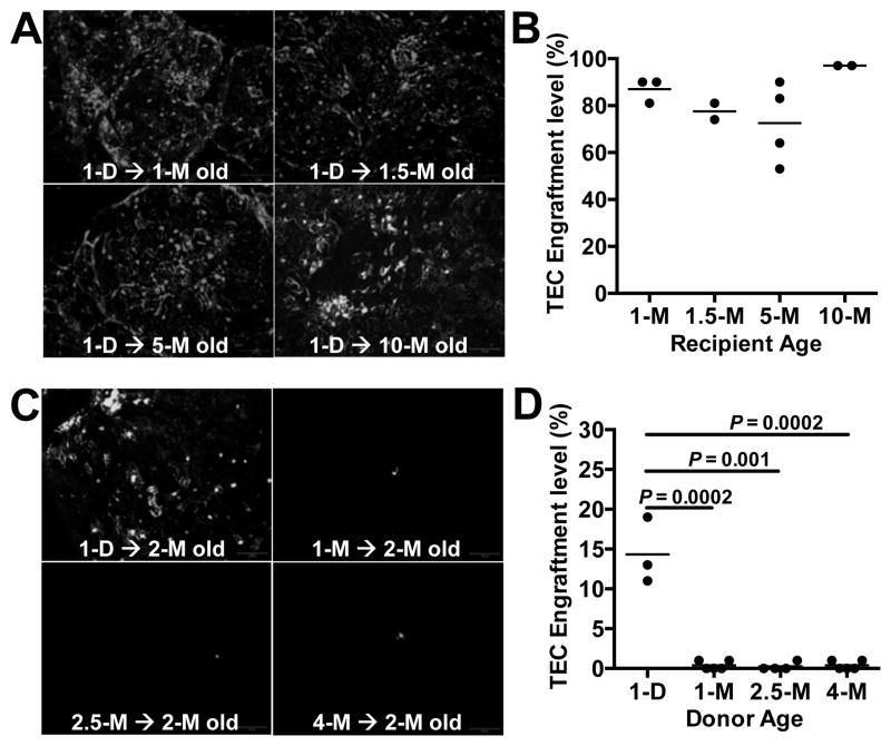
Figure 7.
The thymus retains its ability to be engrafted by TECs regardless of recipient age, but TECs lose their engraftment potential rapidly with donor age. (A) Thymic cells were prepared from P1 GFP-transgenic or RFP-transgenic mice, and equal numbers of EpCAM-enriched TECs were intrathymically injected into 1–10-month old WT mice. After one month, thymuses were PFA-fixed, and completely sectioned (100–200 sections per thymus) and were examined by fluorescence microscopy. Representative sections are shown, and donor cells are shown in gray-scale. (B) Each section was analyzed for the presence of donor cells, and the percentage of sections that contained donor cells within each recipient thymus was determined. Each point on the plot represents the percentage of donor-cell-containing sections within one recipient thymus. Mean values are shown for each experimental group. (C) Equal numbers of EpCAM enriched TECs from GFP-transgenic mice of the indicated ages were intrathymically injected into 2-month old WT mice. After one month, thymuses were recovered and GFP+ cells were detected by fluorescence microscopy. Donor cells are shown in gray scale. (D) Recipient thymuses were analyzed as in (B). Scale bar represents 100 μm. Day and month are shown as D and M, respectively. P values were determined using the unpaired Student’s t-test.

