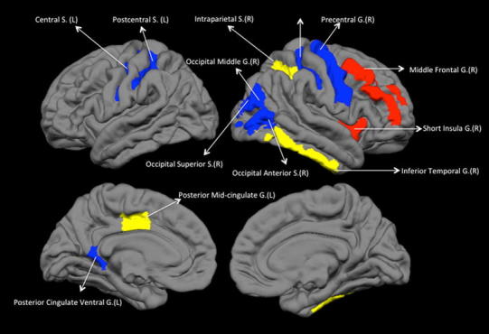
Fig. 2.

Regional changes in topological properties of the gyrification network. A colour figure is provided online. Middle frontal and short insula show increased segregation in patients with schizophrenia (increased in local efficiency/clustering coefficient). Inferior temporal, intraparietal and posterior midcingulate show decreased degree centrality in schizophrenia. All other labelled regions show reduced segregation in patients with schizophrenia. L left hemisphere, R right hemisphere, G gyrus, S sulcus
