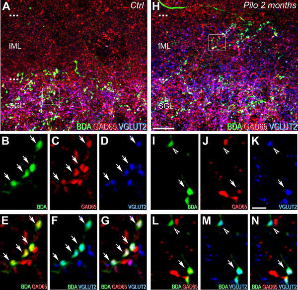
Fig. 3.

Comparison of neurochemical phenotypes for the dorsal dentate gyrus afferents from the SuM between control (a–g) and epileptic pilocarpine-treated (h–n) rats, characterized by simultaneous labeling for BDA anterograde tracer (green), GAD65 (red) and VGLUT2 (blue) in coronal sections. a Image corresponding to a maximum intensity projection of a confocal slice z-stack (22 optical slices, spaced at 285 nm) showing labeling for BDA (green), GAD65 (red) and VGLUT2 (blue) in the dorsal DG of a control rat. Axon terminals and fibers, originating from SuM neurons and labeled for the BDA anterograde tracer (green), were located mainly in the SGL. Numerous GAD65-containing terminals (red) were present in the IML and SGL. VGLUT2-containing terminals (blue) were mainly located in the SGL. b–d Images of the three different fluorophores used for the triple labeling, obtained by sequential acquisition of separate wavelength channels from a single confocal slice in the SGL of the dorsal DG demonstrated that many if not all axon terminals labeled for BDA (b, green, arrows) contained GAD65 (c, red, arrows) and VGLUT2 (d, blue, arrows). e Merge of b and c. f Merge of b and d. g Merge of b–d. Triple-labeled boutons for BDA, GAD65 and VGLUT2 (white, arrows) were surrounded by double-labeled terminals for GAD65 and VGLUT2 (purple) as well as single-labeled terminals for GAD65 (red) or VGLUT2 (blue). h Image corresponding to a maximum intensity projection of a confocal slice z-stack (22 optical slices, spaced at 285 nm) showing labeling for BDA (green), GAD65 (red) and VGLUT2 (blue) in the dorsal DG of an epileptic rat at 2 months after pilocarpine injection. Axon terminals and fibers, originating from SuM neurons, labeled for the BDA anterograde tracer (green) were distributed within the entire IML in contrast to the control rat (compare with panel a). Numerous GAD65-containing terminals (red) were present in the IML and SGL. VGLUT2-containing terminals (blue) were located in the SGL but also in all the IML. i–k Images of the three different fluorophores used for the triple labeling, obtained by sequential acquisition of separate wavelength channels from a single confocal slice, in the IML of the dorsal DG demonstrated two types of BDA-labeled axon terminals (i, green): the first one contained GAD65 (j, red, arrow) and VGLUT2 (k, blue, arrow), the second one contained VGLUT2 only (i–k, arrowhead). l Merge of i and j. m Merge of i and k. n Merge of i–k. Triple-labeled boutons for BDA, GAD65 and VGLUT2 (white, arrow) were surrounded by double-labeled terminals for BDA and VGLUT2 (arrowhead) as well as single-labeled terminals for GAD65 (red) or VGLUT2 (blue). Ctrl control, Pilo 2 months pilocarpine-treated rat at 2 months after status epilepticus. Scale bars 10 µm in a and h; 2 µm in b–g and i–n
