
Fig. 3.
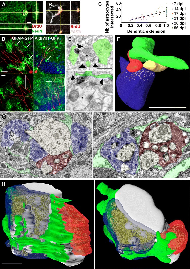
Pre-existing astrocytes ensheathe synapses from adult-born neurons. a, b Confocal micrographs and orthogonal projections of hippocampal sections immunostained for BrdU and NeuN (a) and BrdU and GFAP/S100β (Astro). (b). Scale bar 5 µm. c Scatter plot showing the number of astrocytic territories intersected by newborn neurons, as a function of their normalized dendritic extension (Spearman’s rank correlation test, p < 0.0001, R 2 = 0.64, n = 6–26 neurons per timepoint, 85 neurons in total). d Confocal micrographs of newborn neurons (red) in GFAP-GFP mice (left panels) and Aldh1l1-GFP mice (right panels), illustrating one neuron intersecting the territory of several astrocytes (upper panels, arrows) and several neurons intersecting the territory of the same astrocyte (in white squares, lower panels). Scale bars 20 µm. e Electron micrographs of MSBs (stars) formed by the spine of a newborn neuron (dark immunolabeling) and ensheathed by a perisynaptic process (false-colored in green). Arrowheads point to dendritic spines. f 3D reconstruction of the MSB illustrated in the lower panel of e Scale bars in e and f 0.5 µm. g Electron micrographs of MFT from new neurons (dark immunolabelling, false-colored in red) synapsing with the dendrite or thorny excrescence of a CA3 pyramidal cell (not colorized) that synapse with another non-labeled MFT (blue). The perisynaptic processes are colored in green. h 3D reconstruction of a perisynaptic process (green) ensheathing a MFT from a new neuron (red) and from a non-labeled neuron (blue). Scale bars in g and h 1 µm